TomoTherapy
Accurate, Effective, Customised.
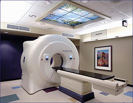
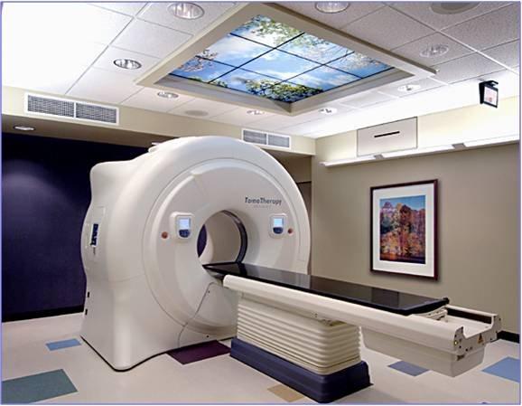
The TomoTherapy® System is a revolutionary radiotherapy system, which redefines the standard for individualized and precise treatment of tumors anywhere in the body — while creating a new paradigm for patient comfort and quality of life. A complete IGRT/IMRT solution, TomoTherapy combines integrated CT imaging for exceptional treatment accuracy with a first-of-its-kind helical treatment delivery platform that uses patented beam-shaping technology to precisely target tumors while minimizing impact on surrounding healthy tissue.
It offers highly accurate 3D dose distribution with a single 360-degree rotation of the linear accelerator, in a very short period of time (~5 minutes), while protecting healthy tissue adjacent to the lesion through a patient-friendly treatment process.
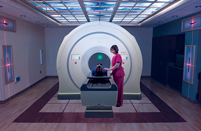
It looks like a spacious CT scanner, while its reliability and ease of handling allow it to simplify even the most complex cases.
Why choose Tomotherapy®
- It uses daily CT images based on the patient's anatomy and the structure of the tumour at the time of treatment, contrary to other methods that use the treatment plan of the previous week or month.
- It irradiates the tumour from all sides with highly precise radiation beams.
- It minimises the possible extension of the radiation to the healthy tissue adjacent to the lesion.
- It adapts to patient needs daily and automatically corrects the treatment plan if necessary.
The Advantages of TomoTherapy
- Increased sparing of healthy tissues adjacent to the treated lesion(s) and minimal treatment toxicity.
- Efficient treatment of elongated targets (e.g., craniospinal irradiation) in a single patient setup process.
- No restrictions on the number, size and shape of treated lesions.
- Accurate treatment delivery through daily volumetric imaging of treated anatomy.
- Online monitoring of treatment delivery on a fraction-by-fraction basis allowing for treatment plan adaptation based on protocol specific action levels.
Total metastases irradiation
When multiple metastases are present, TomoTherapy can target visible tumours in multiple areas of the body with high radiation doses, while limiting damage to healthy tissue.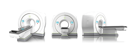 In this case, the doctors use TomoTherapy in combination with chemotherapy, which helps treat smaller metastatic sites, called micrometastases, that may not be visible or treatable only with targeted radiation.
Brain tumours
TomoTherapy allows doctors to treat metastatic brain tumours.
The cancer that has spread to the brain from other parts of the body can be treated with the following steps:
First, the whole brain is treated with a moderately low dose of radiation, not strong enough to damage the brain, but enough to eliminate cancer cells that cannot be seen on scans
Then, the doctors use TomoTherapy to treat all visible tumours by adjusting the radiation, while avoiding damage to healthy cells.
Head & neck cancer
TomoTherapy® makes it possible to treat the lymph nodes in the neck, tongue, oesophagus, and larynx while avoiding injury to the salivary glands. Damage to the salivary glands results in dry mouth.
Being the only radiotherapy standard accepted by the scientific community for the management of complex cases, TomoTherapy® displays a comparative advantage over existing radiotherapy methods and can manage a wide range of cancers such as those of the brain, skull base, neck, spine, liver, spleen, pancreas, kidneys, abdomen, prostate, gynaecological cancers, anal and rectal cancers, sarcomas, lesions of the skin, limbs, breasts, as well as other cancers.
- It can be used in both adults and children.
- It can be used in multiple tumours simultaneously.
- It is a quick, painless procedure that allows patients to return to their daily activities after treatment.
- The entire process is akin to that of a CT scan.
- The main difference compared to other radiotherapy methods, which also constitutes its comparative advantage, is that the daily images from the CT scanner allow accurate repositioning of the patient before each session, and adjustment of the radiotherapy plan in case of anatomical changes of the patient, or shrinking of the tumour.
Prostate cancer
- It uses the computed tomography images to confirm the exact shape and location of a tumour in the prostate, just minutes before the start of treatment.
- It can target even hard-to-reach areas with small, powerful and precise radiation beams in a 360-degree range.
- It minimises treatment-induced side effects in adjacent healthy tissues.
- It avoids exposure to radiation and protects muscle tissue, the spine, lungs and other sensitive organs.
TomoTherapy® promises higher cure rates for patients suffering from cancer.
PreciseART™ - Adaptive Radiation Therapy
DOSE MONITORING, RE-PLANNING, AND DELIVERY VERIFICATION
I. Adaptive radiation therapy (ART)
- During an individual patient’s radiation therapy regimen, adequate dose coverage of the prescribed target volume and dose avoidance for organs at risk (OAR) can be compromised due to interfraction variations in either the target volume or OARs. These variations are patient specific and include translational shifts (e.g., setup error), rotations, and anatomic changes (e.g., tumor shrinkage, organ deformation, weight loss).
- The current standard practice to address interfraction variations is to reposition the patient based on images acquired immediately prior to the radiation therapy delivery (image-guided RT - IGRT). This repositioning method, however, cannot adequately account for interfraction variations such as deformation.
- Adaptive radiation therapy (ART) is introduced to fully address any interfraction variations. ART is a state-of-the-art approach that uses a feedback process to account for patient-specific anatomic or biological changes during the treatment, thus delivering highly individualized radiation therapy for cancer patients.
II. Adaptive radiotherapy integration to TomoTherapy treatments
 The TomoTherapy System combined with the Accuray Precision™ Treatment Planning System and iDMS™ Data Management System, incorporate all necessary tools required for adaptive radiation therapy. These fully integrated systems include: software to inversely plan conformal dose distributions to target volumes, on-board fan-beam megavoltage CT (MVCT) for daily IGRT, Deformable Image Registration (DIR) software to map the daily volumetric MVCT images to the plan CT, and dose calculation and summation software to assess the dose distribution variations during the treatment course due to interfraction anatomic changes. The platform’s unique design with common imaging and treatment isocenter reduces the sources of error, and the fan beam MVCT provides accurate, heterogeneous superposition dose calculation without additional modification or special quality assurance (QA).
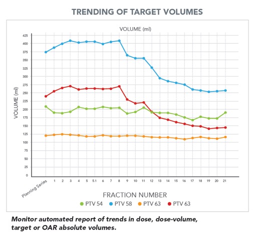 Anatomical or biological changes (e.g., weight loss) of each patient during the radiotherapy treatment regimen are considered using the PrecisesART option.
With PreciseART the doctors monitor the treatment regimen to every patient and efficiently adapt plans helping them to deliver more precise treatments to their patients.
PreciseART:
- Automatically process daily CT images so that clinicians can monitor all patients and set protocol-specific action levels to flag cases for review and possible plan adaptation
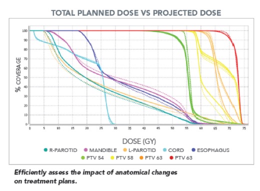
- Offers streamline re-planning capabilities allowing clinicians to efficiently generate new treatment plans based on previous plan data
- Maintain the integrity of original treatment plans to ensure tumor coverage, preserve OAR doses and reduce toxicity
III. Key parameters of PreciseART:
I. Quantitative Images
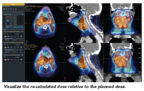 Fan Beam MVCT images allow for treatment-plan-quality dose calculations using the daily IGRT images.
- Accurate, heterogeneous superposition dose calculation
- Automatically augments daily MVCT with superior, inferior and axis aspects from kVCT
- Incorporates daily patient treatment shifts in adaptive calculations
II. Automated Monitoring
Automating key processes allows clinicians to monitor all patients and immediately identify candidates for re-planning.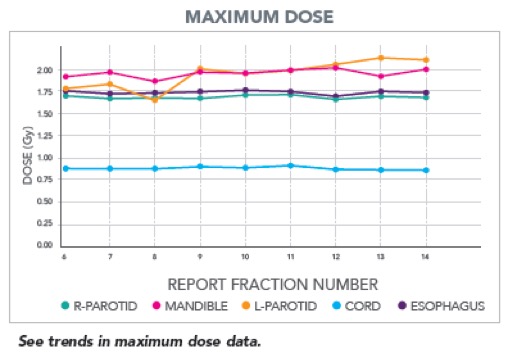
- Deforms the planning VOIs onto the daily image
- Calculates the dose on the daily image
- Deforms and accumulates daily dose onto treatment planning CT
- Generates user-defined reports
- Flags fractions with structure(s) exceeding user-defined dose or dose-volume tolerance
III. Efficient Evaluation
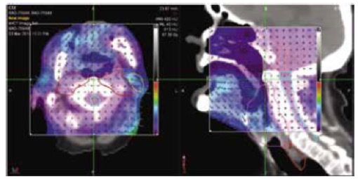 Fully integrated treatment planning and delivery data helps clinicians quickly identify patients that will benefit most from re-planning.
- Review daily dose and registrations, cumulative dose, dose differences and trending data
- Compare fractions and see original and deformed contours on daily merged image
- Evaluate deformation with built-in QA tools
PreciseRTX™ - Making retreatment more efficient and safe
PreciseRTX a workflow-oriented solution
 When treating recurrent disease, the ability to precisely irradiate the target while carefully avoiding healthy normal tissues is essential.
The PreciseRTX™ retreatment software enables the introduction of previous radiotherapy treatments into the new simulation CT in order to be considered during new treatment planning calculations.
The software helps clinicians to accelerate and enhance the process of creating new treatment plans for patients who have received previous irradiation.
PreciseRTX
- Enables importation of patient’s plan data from both Accuray and non-Accuray systems

- Requires no transfer of data back and forth to third-party systems
- Automatically deforms original plan contours onto a new treatment planning CT
- Automatically deforms previously delivered dose onto a new planning CT
- Utilizes information from the original treatment when generating the retreatment plan
- Sums the original and new treatment plans to review the total dose
DELIVERY ANALYSIS™ - Patient specific quality assurance of delivered treatments
I. Delivery Analysis
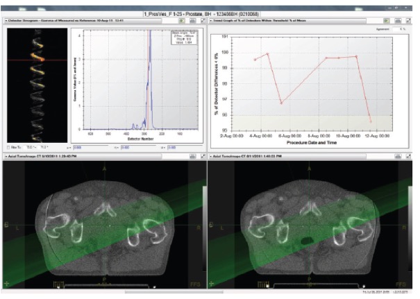
Delivery analysis automatically collects all treatment delivery data and performs a quantitative comparison between planned and measured treatment procedures.
- For every patient, test the multileaf collimator (MLC) using pulse-by-pulse detector signals to measure differences between expected and delivered MLC performance. If inconsistencies exist, the Delivery Analysis software will show both the source of the difference and the potential dosimetric impact to the patient.
- For every fraction, an analysis of exit fluence is generated from exit detector data. Our medical physicists and doctors can easily see if a fraction was not delivered as expected because of possible patient positioning issues, anatomical changes or machine performance.
Intuitive Interface
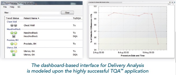
- For all measured data, Delivery Analysis presents data in an easy-to-understand dashboard. This dashboard enables you to see “at a glance” results for all pre-treatment and in-treatment analyses for all patients. Alert levels are configurable by the medical physicist and the clinician.
- For each patient, and for each delivered fraction, the clinician can also review the data in detail. The interface provides four views that can be customized by users to display key information, including: MLC and exit-detector sinograms; CT images with and without calculated dose; MLC leaf histograms; and dose volume histograms (DVHs).
Pre-treatment capabilities
Reconstruct measured MLC delivery pattern via leaf open times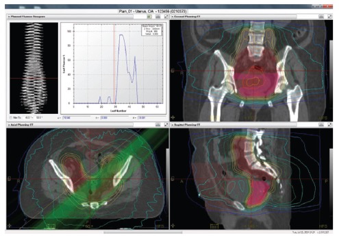
- Calculate dose distribution based on reconstructed MLC delivery pattern
- Automatically analyze and compare planned and measured MLC delivery patterns
- Compare original planned and recalculated dose, including gamma distance to agreement test
In-treatment capabilities
- Compare detector data between selected treatment fractions, both visually and with gamma distance to agreement test
- Analyze and trend differences and other metrics based on detector data for all delivered fractions
- Assess delivery consistency throughout the treatment course
- Visualize treatment delivery for every angle with powerful tools, including ganged images and graphs
