Cardiology
The Cardiology department of Iatropolis Medical Group is fully organized with new digital machine technology and experienced scientific staff, for comprehensive cardiology diagnosis.

The Cardiology Department performs the following diagnostic tests
- Echocardiogram
- Cardiac Stress Test
- Electrocardiogram (ECG)
- Holter Rate/Pressure monitor
- Stress Echo
- Myocardial scintigraphy with a digital γ-camera
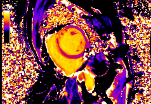
- Cardiac Magnetic Tomography with Myocardial Mapping (T1/T2 Mapping)
- Stress MRI
- CT coronary angiography
Stress Echo
Dynamic echocardiography with dobutamine administration (DSA Stress Echo) is a modern functional ischemia test with high diagnostic value. In fact, in the latest guidelines of the European Society of Cardiology, the stress echo tends to qualify as a first-line functional test for the detection of myocardial ischemia over the electrocardiographic fatigue test (2019 ESC Guidelines for the diagnosis and management of chronic coronary syndromes).
Advantages of the method
The great advantage of the method is its high sensitivity and specificity (>85%) for the detection of significant myocardial ischemia, as well as the ease with which it is performed. The protocol includes administration of dobutamine, lasts less than 30 minutes, and is terminated by administration of an intravenous β-blocker (metoprolol). The patient remains in the room for a few minutes until the dobutamine is metabolized and is safe and then exits and resumes normal activities.
3D real-time visualization
The technique, which is used in our center, includes 3D real-time imaging of the left ventricle in all stages of fatigue. This allows the simultaneous observation in long and short axis (5-7 slices) of the ventricular myocardium, increasing the sensitivity of the method (Assessment of image quality in real time three-dimensional dobutamine stress echocardiography: an integrated 2D/3D approach, Ahmer et al , Echocardiography, 2015). The study is performed with the simultaneous administration of a contrast medium (Sonovue) to improve the image and precisely define the endocardium.
Stress echo indications:
1. Patients with intermediate (15-85%) probability of coronary artery disease for detection of possible ischemia. Such patients are usually over 40 years of age, with at least one classic risk factor for coronary artery disease and chest complaints (typical or atypical).
2. Screening in asymptomatic patients with multiple risk factors for prognosis.
3. For prognosis in coronary patients with a history of acute myocardial infarction, as well as assessment of the effectiveness of medication.
4. Myocardial viability check in patients with prospective coronary reperfusion, as well as for prognosis in patients who are going to undergo surgical or percutaneous aortic valve replacement.
5. Assessment of severity in aortic stenosis (pseudostenosis vs true severe stenosis).
Serious complications (OEM, VT/VF) have an incidence of approximately 1/5000, while that of the classic ECG stress test is 1/1000. Given its higher diagnostic value and the complications of coronary disease prevented, dynamic echocardiography is superior as a functional test. The examination is carried out in the presence of a defibrillator and support equipment in case of serious complications, while the staff is adequately trained (doctor-ALS provider/instructor, nurse-BLS provider).
Overall, 3D dynamic echocardiography is an excellent diagnostic technique for determining the location, severity, and extent of coronary artery disease, and has applications in severe valvular disease.
Myocardial scintigraphy with a digital γ-camera
The first digital imaging system of myocardial perfusion in the Balkans, with the D-SPECT digital γ-camera, was installed and is operating in the diagnostic unit of Patisia, performing all myocardial scintigraphy.
For more information
Cardiac Magnetic Tomography with Myocardial Mapping (T1/T2 Mapping)
These are newer techniques, which are applied in the Iatropolis Medical Group. Mapping assigns a T1 or T2 time number to each image point (voxel) forming a digital map. This results in the "objectification" of the image, without its interpretation depending on the subjective diagnostician. Thus, there are limits of normal values, beyond which pathology is diagnosed. This technique dramatically increases the diagnostic value of the test by detecting invisible myocardial damage.
Examination Protocol
A classic protocol applied in the majority of cases takes 35-40 minutes, a time easily tolerated by the average patient. Additional sequences, such as Stress MRI, extend the examination, no more than 10 minutes. In addition, venipuncture is needed to administer the contrast agent.
Diagnostic Value
Magnetic resonance imaging of the heart provides a multitude of tools to reveal most diseases of the cardiovascular system without dangerous ionizing radiation, administration of toxic contrast agents (gadolinium is also administered to patients with renal failure, while it is practically devoid of side effects) and with unique diagnostic accuracy.
Stress MRI
This technique has a very high sensitivity, higher than stress echo, for the detection of myocardial ischemia and is compared to FFR (fractional flow reserve), in terms of detecting patients with significant coronary disease (Magnetic Resonance Perfusion or Fractional Flow Reserve in Coronary Disease, Nagel et al, NEJM, 2019). In addition, it is perhaps the best technique for revealing microvascular coronary disease, which is underdiagnosed due to a lack of reliable imaging techniques.
First-pass perfusion: This sequence is used in stress MRI after injection of a vasodilator. The loading agent we use is regadenosone 400 mg (Rapiscan), which is bolus-infused at the same dose, regardless of body weight. It is a safe agent, with minimal side effects, it does not cause real ischemia like the classic fatigue test or stress echo, and it also has an antidote (aminophylline).
CT coronary angiography
CT angiography is a new and bloodless examination for the control of the coronary vessels. The examination first appeared with the creation of the sophisticated polytomous CT scanners. The exam is very simple and no preparation is required. Its time duration is about 10 minutes and the "slices" are taken by holding a breath for 10-15 seconds.
An intravenous drug is administered to delineate the coronary vessels and the patient can return to his daily activities after the examination. Considering that coronary artery disease is now the leading cause of morbidity and mortality in economically developed countries, it seems that CT angiography is now a routine examination.
Calcium Scoring
The Calcium Scoring measurement measures and evaluates calcium deposition in the coronary vessels of the heart and is an important indicator of the possibility of developing serious heart disease, or even strokes. Calcium deposits in the arteries of the heart are a strong sign that atherosclerotic plaques are likely also present within the coronary vessels, capable of leading to heart attack and/or sudden death.
Population groups for which the examination is indicated
- Patients with atypical angina pectoris
- People with high predisposing factors for coronary heart disease
- By-pass patients who will be able to avoid serial catheterizations to check their grafts
The following must be emphasized: people with atypical angina pectoris or with high predisposing factors are a population that does not have diagnosed coronary artery disease. In this population CT coronary angiography functions more as a preventive control (screening) to diagnose a serious condition in time.
Its sensitivity and specificity are quite high and even higher than those of other tests used for preventive screening such as mammography.
It is the safest, painless, bloodless and non-traumatic screening method for all categories of examinees and patients, because it does not show any of the possible complications like classic coronary angiography.
At the same time, it is an extremely simple examination in its execution process and its cost is much lower than that of classic coronary angiography.
Coronary angiography at Iatropolis Medical Group
In the Iatropolis medical group, the examination has been carried out for fifteen years, and there is an ever-increasing interest in it from the medical world. In the first steps there were several problems, which had to do with two characteristics of the vessels of the heart: their small dimensions and their constant movement with the heartbeat. Patients with more than 65 contractions per minute could not be tested reliably. Gradual improvements occurred in the intervening period, but the problem of vascular movement was not finally overcome. Thus a significant number of examinations were non-diagnostic or in one examination parts of the vessels could not be assessed. These limitations delayed the acceptance of CT angiography by the cardiology community and its wider application.
The new innovative Siemens SOMATOM Definition Flash CT has the innovation of carrying two detector lamp systems. With this technology the time needed to create an incision is 83 milliseconds, exactly half compared to the machines of the immediate previous generation. The experience of the Iatropolis group in a large number of tests carried out in recent years shows that the movement of the coronary vessels in patients with high heart rates is no longer a problem.
At the same time, the results from publications in reputable medical journals show very high accuracy compared to classic coronary angiography.
The negative diagnostic value of the test (NPV) is 99.4%, which means that, if it is normal, the patient does not suffer from coronary artery disease.
The positive diagnostic value (PPV) is 85.7% which means that if there are findings on CT coronary angiography, the patient probably has significant stenoses of their coronary vessels.


