PET/CT
BIOGRAPH VISION
BIOGRAPH mCT Flow
Three (3) unique PET/CT systems for the first time in Greece - The latest technology in molecular imaging
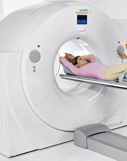
PET/CT imaging offers us early diagnosis of cancerous tumors, or their recurrence as well as other pathological conditions. The BIOGRAPH VISION and BIOGRAPH mCT Flow systems are at the forefront of PET/CT molecular imaging examinations. Their advantages are the following:
- Maximum examination speed, minimum radiation dose.
- Even more detailed visualization of each instrument.
- Ability to detect even the smallest tumors.
- Comfortable examination for all patients regardless of body type.
- Digital technology and application of artificial intelligence.
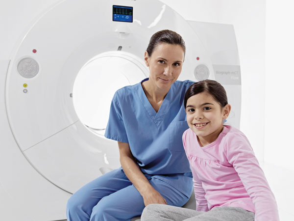
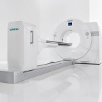
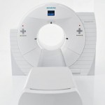
The tests are carried out by specialized staff and evaluated by doctors with great experience and expertise in the method. The doctors who staff the PET/CT department are Dr. Lachanis Stefanos (Director Radiologist), Dr. Karantanis Dimitrios (Director Nuclear Physician), Dr. Vassilis Voliotopoulos (Scientific Lead Nuclear Physician), Dr. Kalkanis Dimitrios (Nuclear Physician), Dr. Loukas Lambrakos ( Nuclear Physician) and a team of medical radiologists and nuclear physicians.
Leading the Way in Prostate Cancer Diagnosis – IATROPOLIS Group
The Oncology Clinic of IATROPOLIS is proud to be the first in Greece to offer the advanced PSMA PET/CT scan, setting a new standard in accurate diagnosis and staging of prostate cancer.
With state-of-the-art PET/CT technology and a team of specialized medical professionals, we provide patients with unmatched diagnostic precision and speed.
What Makes PSMA PET/CT Special
PSMA PET/CT (Prostate-Specific Membrane Antigen PET/CT) is a revolutionary imaging test that detects prostate cancer at the molecular level.
By targeting PSMA, a protein highly expressed in prostate cancer cells, it can locate even the tiniest cancer lesions with exceptional accuracy.
Benefits for Patients
- Pinpoints metastases or recurrence with incredible precision
- Enables timely and targeted treatment plans
- Non-invasive and completely safe
- Quick procedure with immediate results
- Reduces the need for unnecessary treatments or surgeries
The PSMA PET/CT scan is fully covered by the Greek public health insurance fund E.O.P.Y.Y (Ε.Ο.Π.Υ.Υ) , giving all insured patients access to this cutting-edge diagnostic method at no extra cost.
IATROPOLIS – At the Forefront of Innovation
At IATROPOLIS, we continue to lead advancements in cancer imaging and treatment, combining innovation, quality, and compassionate care to support every oncology patient.
For more information or to schedule your PSMA PET/CT, contact our Oncology Clinic at 2106796000.
What is a PET/CT examination
Positron Emission Tomography (PET) is an imaging test that provides the possibility of detecting any pathological conditions at an early stage, because it depicts biochemical changes, which manifest themselves long before the anatomical ones, which are depicted by conventional imaging methods.
It is the most modern imaging method of nuclear medicine with a wide range of clinical applications in Oncology, Cardiology and Neurology.
When to have a PET/CT
PET/CT offers an effective way to examine the metabolic activity of the human body and is mainly used in cancer patients.
Cancer cells, due to their high metabolic rate, are visualized as more intense "bright" spots in the PET scan, as a result of which, diseased areas can be identified more easily.
Thus, the PET scan is used to determine the extent of the disease, or how well the malignant disease is responding to treatment, or finally to determine whether a treated malignant disease has recurred or not.
Your doctor, who asked you to take this test, will use its result to make very important decisions, which usually have to do with choosing the necessity but also the type of surgery, chemotherapy, radiation therapy, or even simple follow-up at regular intervals.
The quality of the examination and its accurate interpretation are therefore of vital importance.
Preparation of PET/CT examination
Detailed preparation instructions are given to you at the time of your appointment. Some general rules that apply to your preparation are as follows:
- Fast for at least 6 hours before your appointment. During this time you are only allowed to drink water (no coffee or soft drinks). Even a small amount of food can significantly degrade the reliability of the test.
- You can take your medicines normally (with water for drinking), except for sugar medicines, for which you will be given special instructions when we contact you.
- Women of childbearing age should make sure they are not pregnant.
- Avoid vigorous physical activity (exercise) and exposure to cold temperatures for 24 hours before the test.
- It is a good idea to wear comfortable clothes when you come for the examination. Avoid metal buttons, zippers, belts, or metal objects.
- Collect and take with you all your Medical file. Some information may be very important to the interpretation of the test you are about to take.
- Before starting your appointment, fill out any information or forms that were requested when you booked your appointment.
PET/CT Examination
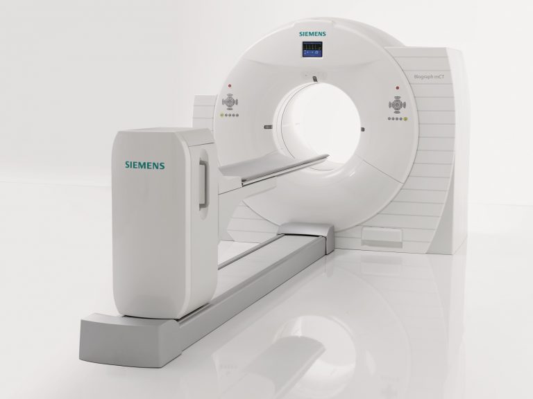
The machine on which you will be examined is very similar to a computed tomography (CT) scan.
On the day of your appointment, after being greeted by the receptionist, you will be asked some simple questions about your medical history.
Your blood sugar will then be measured, the test procedure will be explained to you in detail, and the radiopharmaceutical (18-FDG) necessary for the test will be given intravenously. The administration of the drug is painless and does not involve any risk of an allergic reaction.
After the radiopharmaceutical has been administered, you will lie comfortably in a quiet, specially designed area for about an hour, at the end of which you will be asked to urinate (to empty your bladder) and then you will be transferred to the special bed PET/CT camera.
There the specialized staff will place you in a suitable position and ask you to remain as still as possible in order to take the necessary images.
The acquisition of the images is painless and harmless to the patient and is performed in a single scan lasting 8-12 minutes with a significant reduction in radiation thanks to the FlowMotion technique and improved imaging quality through the OncoFreeze AI software.
The personalized anatomy application provides personalized imaging, precisely based on the examinee's anatomy, tailored precisely to their needs. The application of A.I. (Artificial Intelligence), provides additional reliability in the diagnosis of the test.
When the test is over and it has been checked for completeness you will receive some very simple instructions and you can then leave the clinic.
BIOGRAPH mCT & BIOGRAPH VISION
The BIOGRAPH mCT Flow and BIOGRAPH VISION systems are an innovation in the traditional PET/CT method. Conventional PET/CT systems perform the examination through a process of successive movements of the examination table and the patient's position. This results in a higher radiation exposure to the patient, a lower resolution of the exam, and increased patient stress from pausing and restarting the exam.
The BIOGRAPH mCT Flow and BIOGRAPH VISION systems eliminate the above unpleasant consequences, due to their innovative design and the digital technology they use. An examination lasting only 8-12 minutes detects a wide range of cancers such as breast, oesophagus, cervix, melanoma, lymphoma, lung, rectum, brain, SCS, ovary, liver, etc. In addition, it is used in the diagnosis and study of prostate cancer, the disease Alzheimer's as well as in the study of myocardial viability.
The advantages of the PET/CT examination with the BIOGRAPH mCT Flow and BIOGRAPH VISION systems are the following:
- Even more accurate functional and anatomical imaging
- Diagnosis at an early stage
- Accurate localization and staging of the disease
- Re-staging
- Ability to detect even the smallest tumors
After the PET/CT scan
Generally there are no special restrictions after the examination. It is advised to avoid prolonged close contact with pregnant women and small children for 6 - 8 hours.
You can eat normally and return to your normal physical activity immediately after your test.
It is advised to drink a little extra water for the rest of your day.
Accurate imaging of every organ
With FlowMotion technology, parameters such as image resolution, examination speed and management of organ movements due to respiratory function can be incorporated into a test protocol that is adapted to the imaging of every organ.
As a result, the referring clinician receives better resolution images and more accurate SUV quantifications for every organ, leading to more accurate diagnostic results.
The FlowMotion technology maximises image quality, providing imaging protocols based on the needs of the examined organs. It provides integration of variable speed, motion management and a 400x400 image reconstruction matrix into a single scan protocol, based on the needs of each organ and patient. This achieves the finest detail for every organ and enables a more reliable diagnosis.
With old generation PET/CT technology, small or low-grade lesions can go undetected or may not be classified clearly, thereby lowering the predictive value of the examination.
The Biograph mCT Flow system overcomes the above limitations by providing excellent image resolution, with a single patient scan, without compromising the efficiency of the PET/CT examination.
Using the FlowMotion system, a new standard in PET/CT imaging is introduced.
Minimum Dose and Maximum Speed
PET/CT scanners are equipped with the following subsystems: TrueV, which increases the count rate performance of the PET examination up to 2 times. CARE, which dynamically changes the data of the CT scanner lamp, reducing the dose of radiation during the axial tomography (CT) examination, without degrading the image quality.
To minimise the dose to the patient, the scanner allows physicians to visualize only the regions of interest, thus limiting irradiation to the rest of the body, and can complete a whole-body PET/CT examination in just 5 minutes.


